
|
|
Differential adhesion controls columnar unit formation |
| |
Columnar structure is a basic unit of the brain, but the mechanism underlying its development remains largely unknown. The medulla, a part of the visual center in the fly brain, provides a unique opportunity to reveal the mechanisms of three-dimensional organization of the columns. In this study, using N-cadherin as a marker, we revealed the donut-like columnar structures along the two-dimensional layer in the larval brain that evolves to form three distinct layers in pupal development. Column formation is initiated by three core neurons, R8, R7, and Mi1, which establish distinct concentric domains within a column. We demonstrated that N-cadherin-dependent relative adhesiveness of the core columnar neurons regulates their relative location within a column along a two-dimensional layer in the larval brain according to the differential adhesion hypothesis.
|
|
|
|
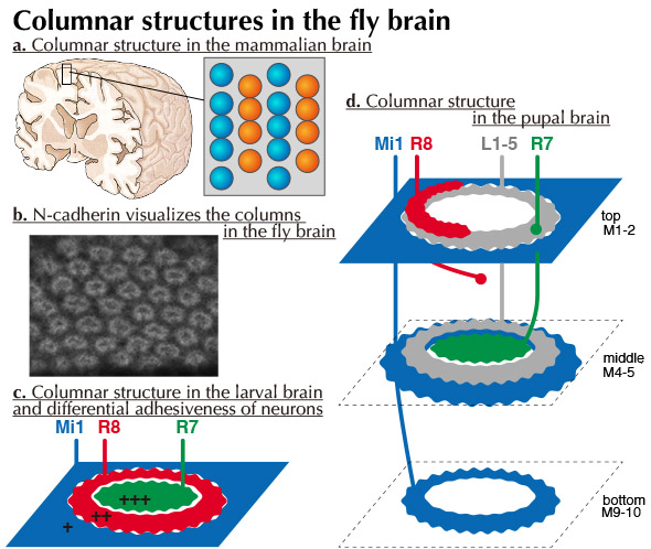
The columnar unit is a basic feature of the brain. However, the developmental mechanisms underlying columnar unit formation remain unclear (Fig. a).
We showed that the cell adhesion molecule N-cadherin reveals a donut-like structure of the column in the fly brain (Fig. b). The two-dimensional donut-like pattern in the larval brain evolves to establish the three-dimensional pattern in the top, middle, and bottom layers during pupal development (Fig. c-d). We identified three core columnar neurons essential for column formation in larval development. The terminal of R7 occupies the dot-like central region of the larval column. The R8 terminal enwraps the R7 terminal forming a donut-like region that overlaps with the N-cadherin pattern. The Mi1 terminal occupies a grid-like region outside the R8 terminal. Thus, R7, R8, and Mi1 form the two-dimensional concentric organization composing the three distinct domains in the larval brain (Fig. c).
The differential adhesion hypothesis proposes that mixtures of cell populations segregate according to their adhesive properties: the less-adhesive cell population is located at the periphery of the cell aggregates and surrounds the more-adhesive cells. Interestingly, we demonstrated that the core columnar neurons express different levels of N-cadherin in the order of R7>R8>Mi1 (Fig. c).
|
|
|
| |
|
| |
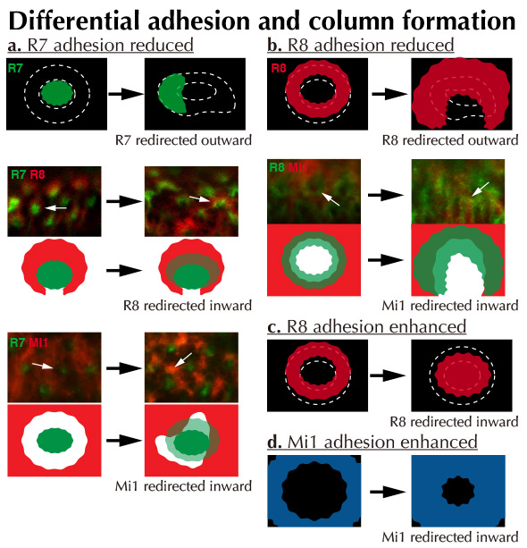
We established a series of genetic tools that can specifically express fluorescent markers and manipulate N-cadherin activity in R7, R8, and Mi1. By using these tools, we clearly demonstrated the in vivo evidence of differential adhesion hypothesis among R7, R8, and Mi1 in column formation in the fly brain.
When the adhesiveness of R7 is reduced, the R7 axon terminals were redirected toward the column periphery (Fig. a). At the same time, the terminals of R8 and Mi1 were redirected toward the column center. When the adhesiveness of R8 is reduced, the R8 axon terminals were redirected toward the column periphery (Fig. b). At the same time, the terminals of Mi1 were redirected toward the column center. When the adhesiveness of R8 and Mi1 is enhanced, their axon terminals were redirected toward the column center (Fig. c-d). There results support the idea that the differential adhesiveness of R7, R8 and Mi1 establishes the concentric organization of the columnar structure.
|
|
|
| |
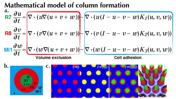
However, it is still unclear if differential adhesion is sufficient to explain the concentric organization of the columns. We therefore established a mathematical model of column formation, in which differential adhesiveness and volume exclusion between R7, R8 and Mi1 are considered (Fig. a). The results of computer simulation demonstrated that the concentric organization of the columns can be reproduced if the relative adhesiveness of the core columnar neurons are preserved (Fig. b-c). Thus, differential adhesion could be the major driving force in establishing the columnar structure.
|
|
|
Column formation is an unexplored important research field in neuroscience. In this study, we identified and developed many essential tools to investigate molecular mechanisms of column formation using the fly brain as a model system. There are a lot of molecules and neurons that play essential roles in column formation. We would like to expand this research to clarify evolutionarily conserved mechanisms of column formation by combining molecular genetics, imaging techniques and mathematical modeling.
Trush, O., Liu, C., Han, X., Nakai, Y., Takayama, R., Murakawa, H., Carrillo, J. A., Takechi, H., Hakeda-Suzuk, S., Suzuki T. and Sato, M.
N-cadherin orchestrates self-organization of neurons within a columnar unit in the Drosophila medulla.
Journal of Neuroscience 39, 5861-5880 (2019).
Carrillo, J. A., Murakawa, H., Sato, M., Togashi, H. and Trush, O.
A population dynamics model of cell-cell adhesion incorporating population pressure and density saturation.
Journal of Theoretical Biology 474, 14-24 (2019). |