


|
|
Developmental mechanisms that create functional neuronal circuits
in the Drosophila visual center
 |
| |
The Drosophila visual center shares many important features of neurogenesis with mammalian brains: production of wide variety of neurons in accord with order of neuron production and formation of layer and columnar structures accompanying neuronal migrations. Additionally, powerful neurogenetic tools of Drosophila allow us high-resolution functional analyses of neuronal circuits. Using the Drosophila visual system as a model, conserved mechanisms that govern formation of functional neuronal circuits will be elucidated.
|
|
| Development of the cerebral cortex |
|
| |
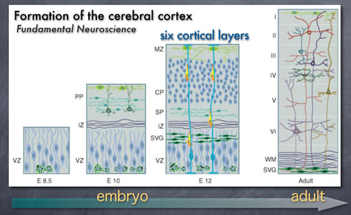
During the formation of the cerebral cortex during mammalian brain development,
neurons migrate from inner to outer areas of the brain to form six cortical layers.
The process of layer formation during embryogenesis has been extensively studied by many researchers. However, it is technically very difficult to relate developmental origin and function of a neuron in the mammalian brain.
|
|
|
| The visulal center in the fly brain |
|
| |
Therefore, we use Drosophila visual center as a model.
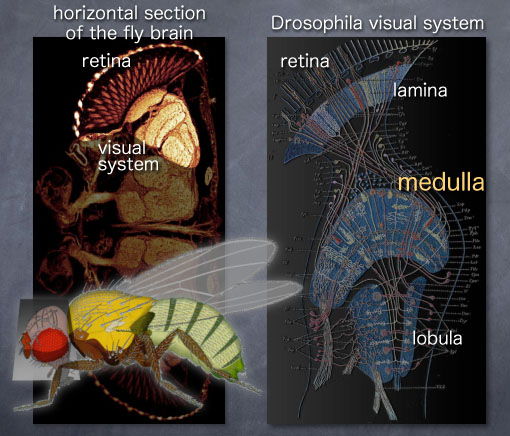
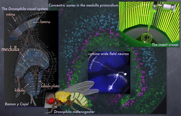
This is a horizontal section of the visual system in the brain.
Visual information received in the retina is transmitted through the structures called lamina, medulla, lobula and lobula plate. We are especially interested in the largest part called the medulla.
The medulla is comprised of 40,000 neurons of at least 60 types forming 10 layers and 800 columnar units. There are many structural characteristics shared by mammalian brains. So, the medulla may model neuronal circuits in the mammalian systems. However, development and function of the medulla remain mysterious.
|
|
|
| Development of Drosophila |
|
| |
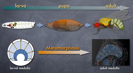
As you know, flies grow through larva, pupa and adult.
The adult medulla shown in the bottom right is already prepared in the larval brain.
During metamorphosis, the larval medulla is significantly modified to form the mature adult structure.
|
|
|
| Concentric zones in the larval medulla primordium |
|
| |
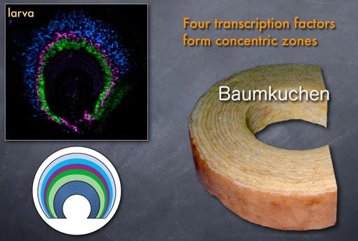
However, almost nothing has been known about developmental mechanisms that pattern the larval medulla. We therefore searched for genes that show interesting expression pattern in the larval medulla. As a result, we found four transcription factors that show concentric expression pattern.
In the photos shown above, only three of them are shown: green, magenta and blue.
Their expression domains are complementary to each other,
suggesting that the developing medulla is similar to a kind of cake, baumkuchen.
|
|
|
|